









.webp)





















Overview
- The major salivary glands are paired organs and include the parotid, submandibular, and sublingual glands.
- They are compound tubuloacinar exocrine glands.
- Each gland is surrounded by a thin capsule.
- The capsule extends septa into the glandular parenchyma, dividing it into lobes and lobules.
-
Functions :
- Produce and secrete saliva into the oral cavity.
- Maintain hydration and protect the oral mucosa and teeth.
- Lubricate the oral cavity during speech, mastication, and swallowing.
- Begin carbohydrate digestion through salivary amylase.
- Contribute to oral immune defense through antimicrobial components, including secretory IgA.
-
Parotid Gland :
- The parotid glands are the largest major salivary glands, located in the parotid region, anterior and inferior to the ear.
- The main parotid duct is the Stensen duct. It opens into the oral cavity opposite the upper second molar.
- May contain intraparenchymal lymph nodes.
-
Submandibular Gland :
- The submandibular glands are also called submaxillary glands, located beneath the mandible, in the submandibular region, with a deep portion extending toward the floor of the mouth.
- The main submandibular duct is the Wharton duct. It opens at the sublingual caruncle, just lateral to the lingual frenulum.
-
Sublingual Gland :
- The sublingual glands are the smallest major salivary glands, located in the anterior floor of the mouth, beneath the oral mucosa.
- Numerous small ducts of Rivinus drain directly into the floor of the mouth along the plica sublingualis.
- A larger sublingual duct, the Bartholin duct, may join the submandibular duct near the sublingual caruncle.
- Microscopically, the functional structure of a salivary gland consists of secretory acini and a duct system.
Acini
-
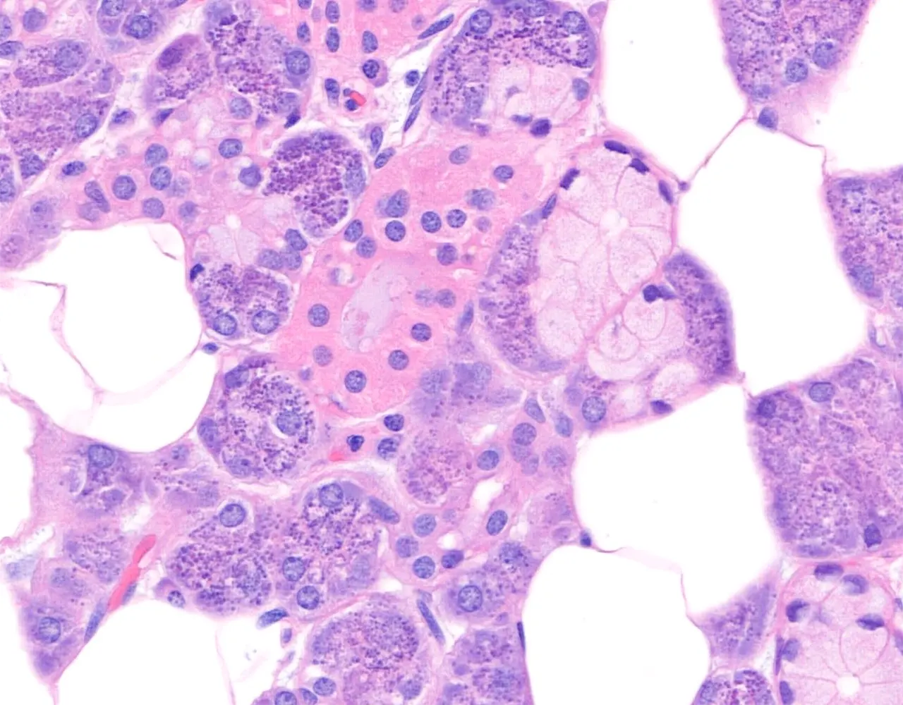
Each gland has a different acinar composition :
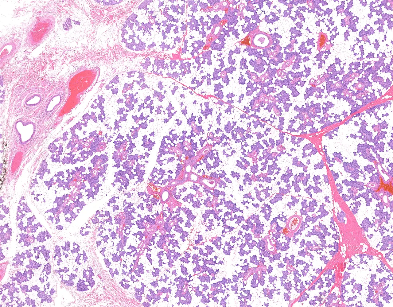
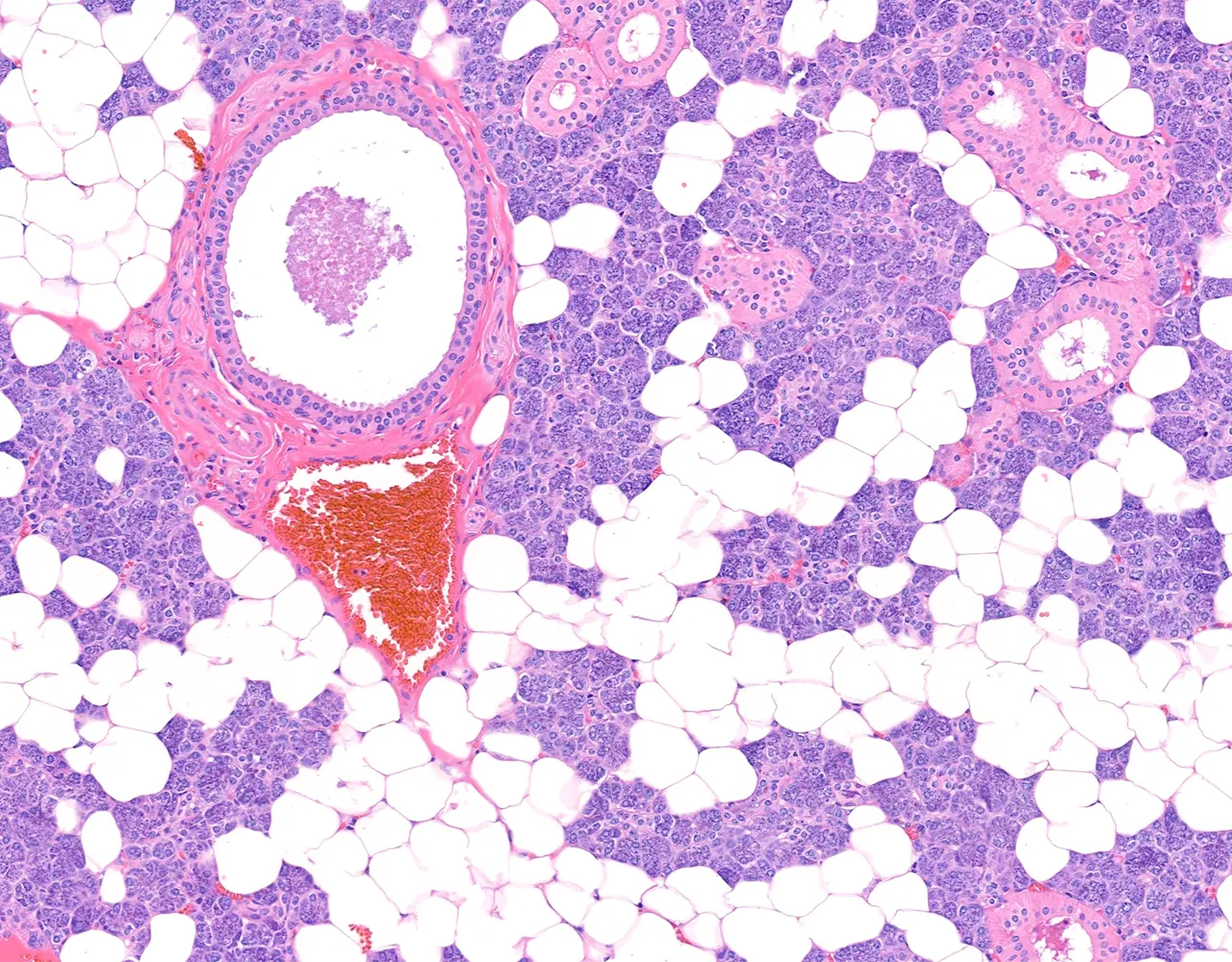
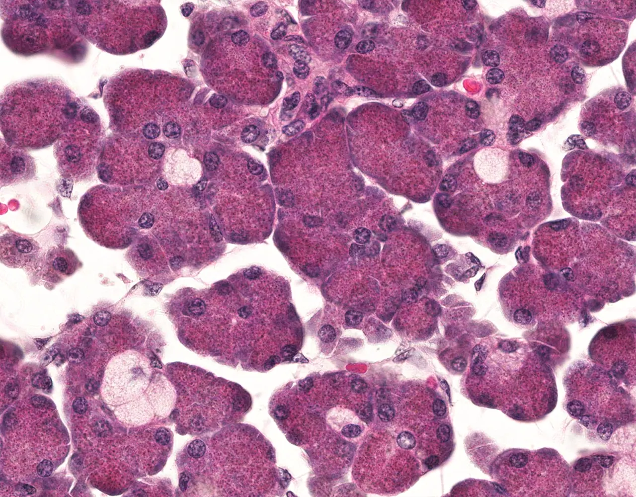
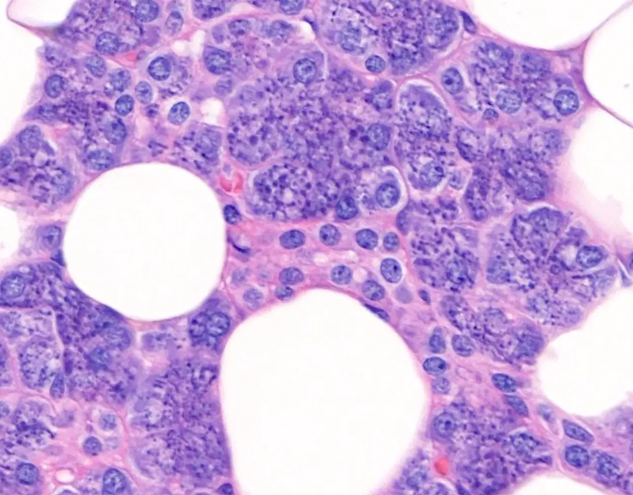
- The parotid gland is almost entirely serous.
- The submandibular gland is mixed, with serous predominance.
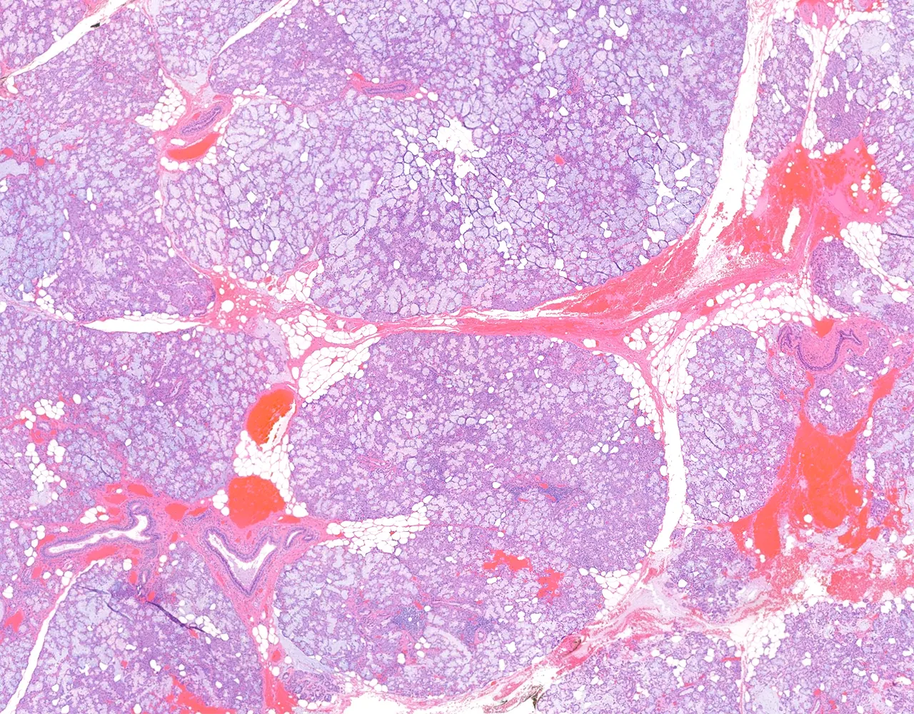
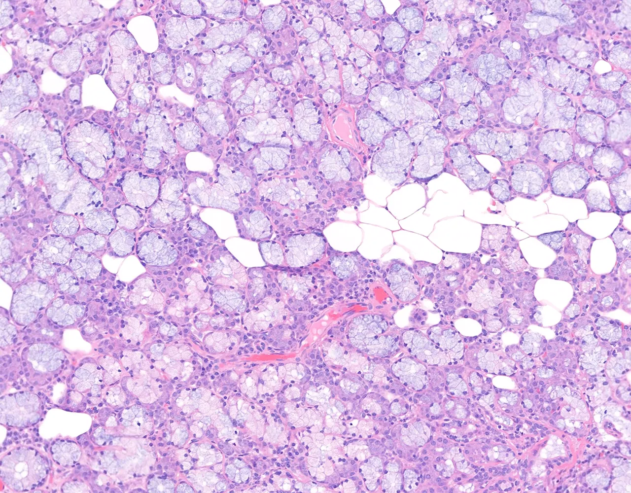
- The sublingual gland is mixed, with mucous predominance.
-
Serous Acini :
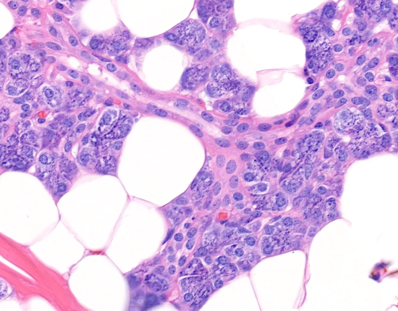
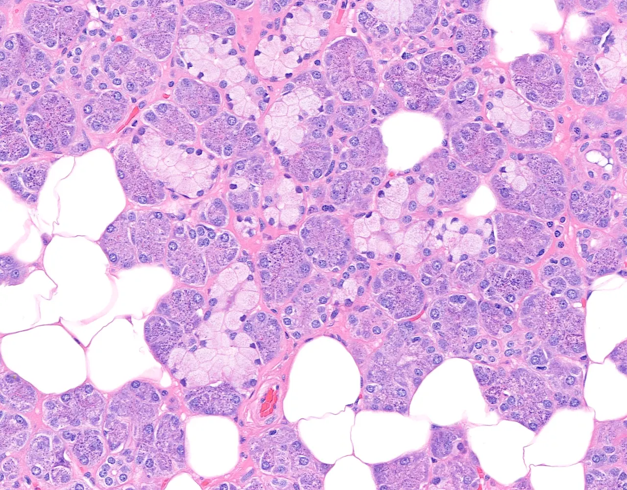
- Serous acini are usually rounded or spherical.
- They are composed of pyramidal serous cells with round nuclei, usually located basally.
- The basal cytoplasm is basophilic because of abundant rough endoplasmic reticulum.
- The apical cytoplasm contains eosinophilic zymogen granules.
-
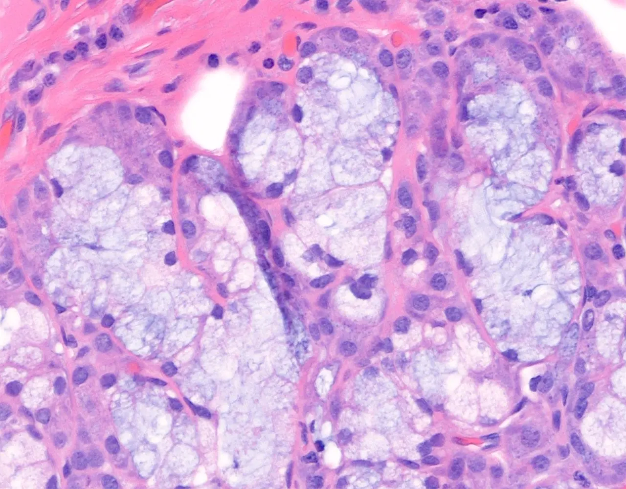
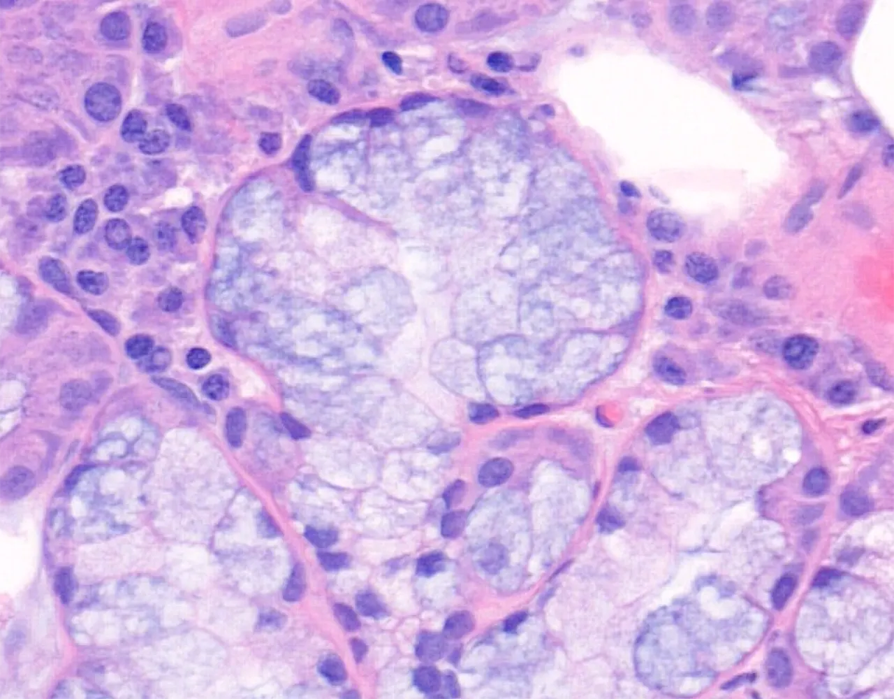
Mucous Acini :
- Mucous acini are usually tubular or tubuloacinar.
- They are composed of pale mucous cells with flattened nuclei displaced toward the basal part of the cell.
- The cytoplasm contains mucin-rich secretory material.
-
Mixed Acini :
- Mixed acini contain both mucous cells and serous cells.
- The mucous cells usually form the main portion of the acinus.
- Serous cells may appear as crescent-shaped caps, known as serous demilunes or crescents of Giannuzzi, at the periphery of mucous acini.
-
Myoepithelial and Basal Cells :
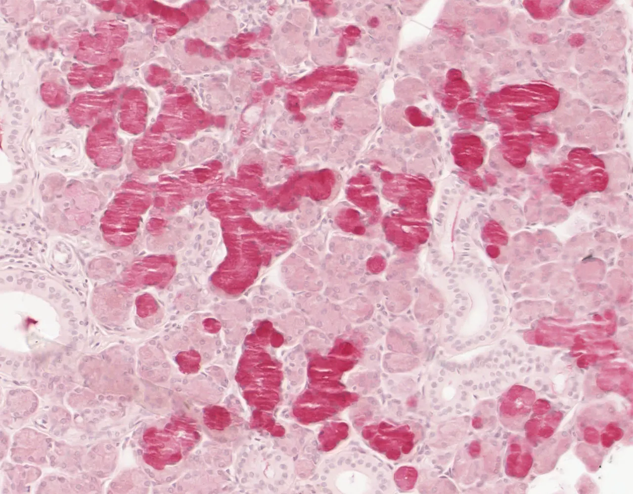
- Myoepithelial cells are flattened contractile cells that are difficult to identify on routine H&E staining.
- They are located between the secretory epithelial cells and the basal lamina, mainly around acini and intercalated ducts.
- Their contraction helps expel secretory products into the duct system.
- Basal cells are noncontractile cells present mainly in larger ducts, especially excretory ducts.
Duct System
- The duct system carries saliva from the acini to the oral cavity.
- The duct system is branched and includes intercalated ducts, striated ducts, interlobular ducts, and main excretory ducts.
-
Intercalated Ducts :
- Intercalated ducts are intralobular ducts, located between the acini and the striated ducts.
- Lined by low cuboidal epithelium and have a usually narrow lumen.
-
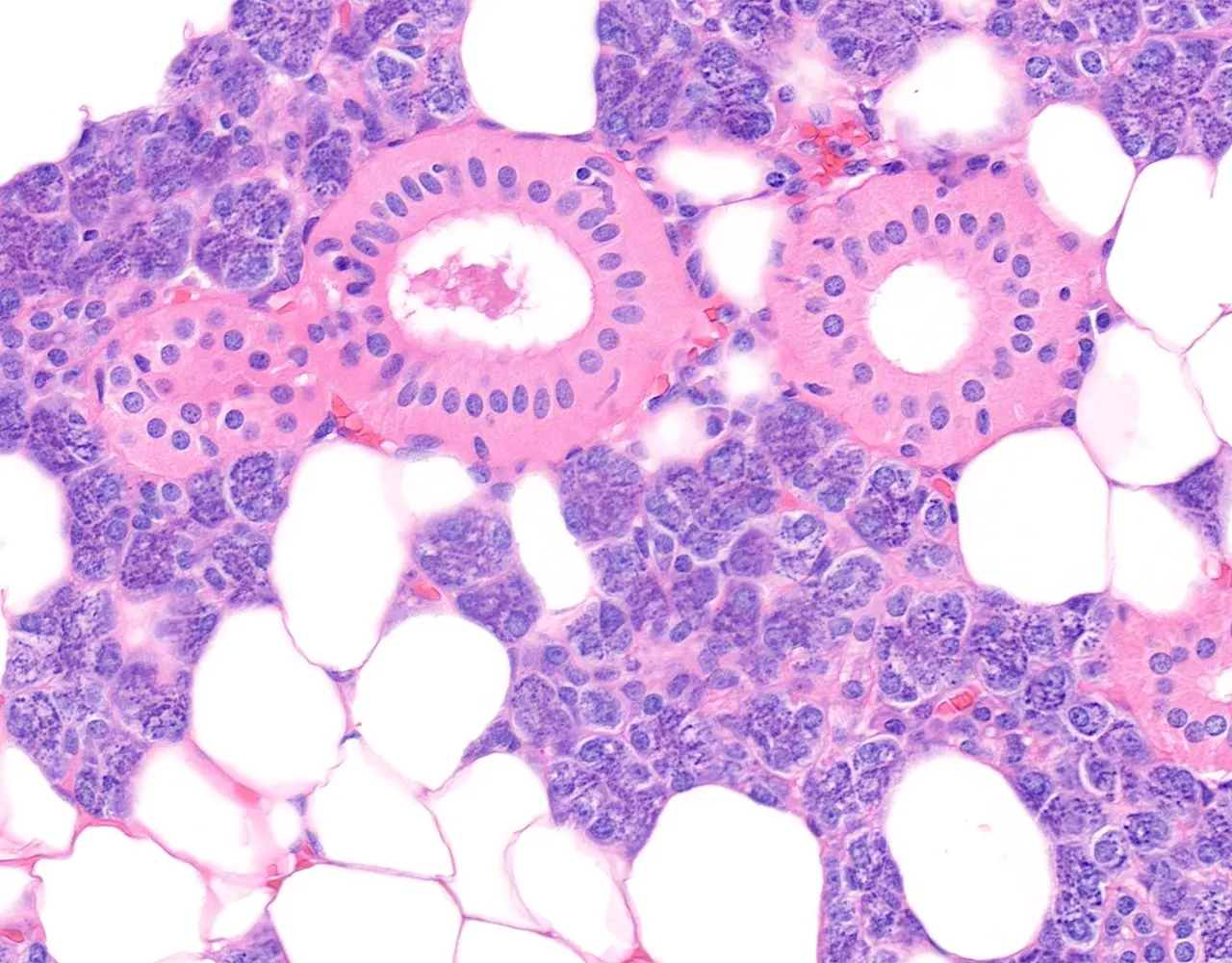
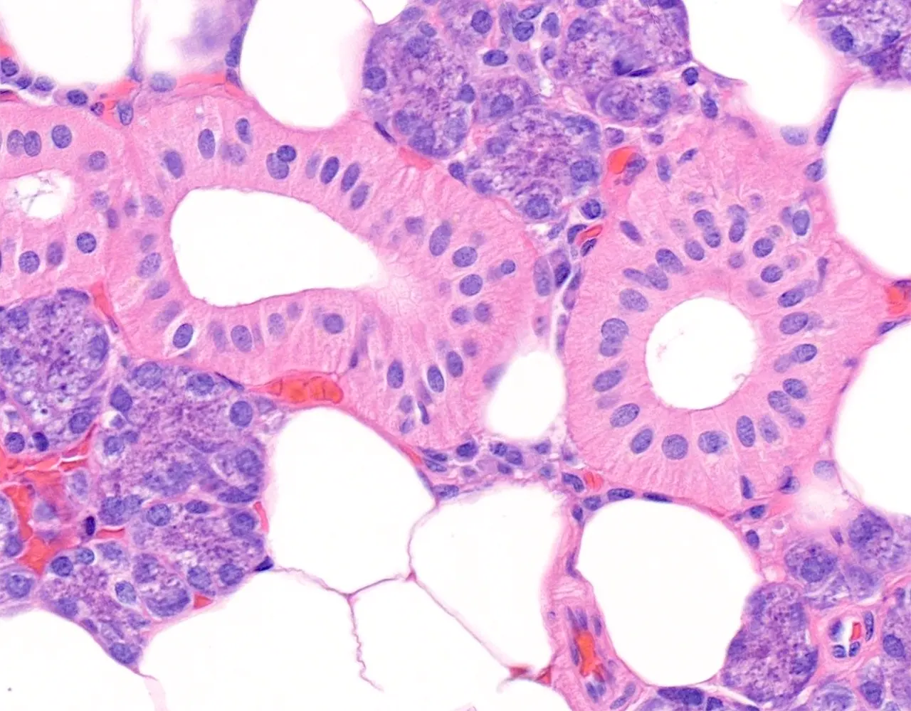
Striated Ducts :
- Striated ducts are also intralobular ducts, located between the intercalated ducts and the larger excretory ducts.
- Lined by simple cuboidal to columnar epithelial cells with eosinophilic cytoplasm.
- Basal striations are visible at the base of the cells.
- These striations correspond to basal plasma membrane infoldings.
-
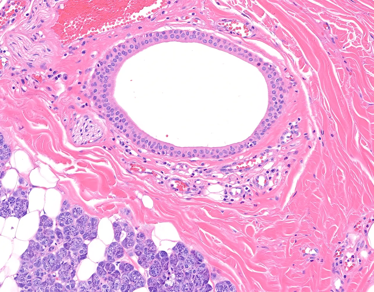
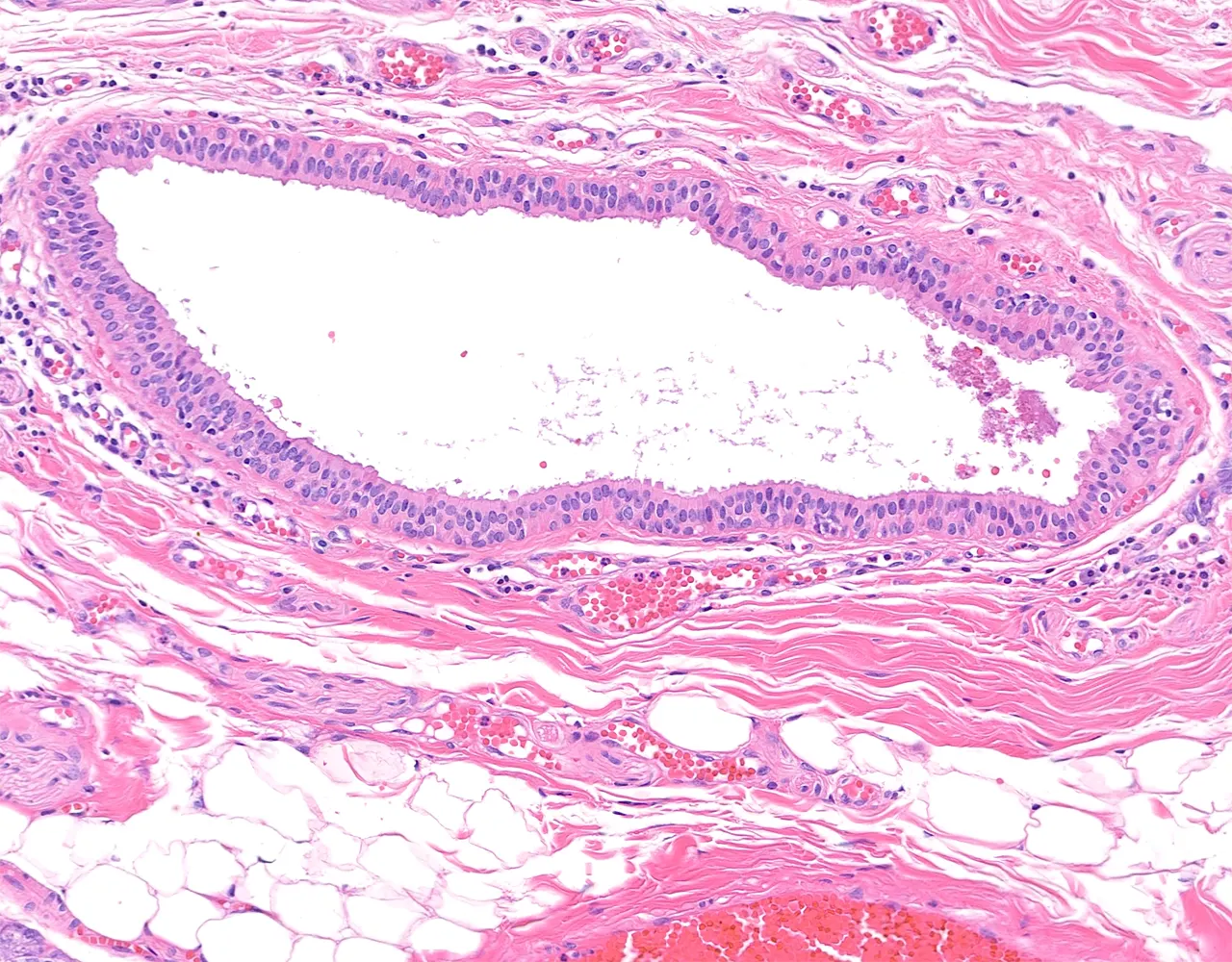
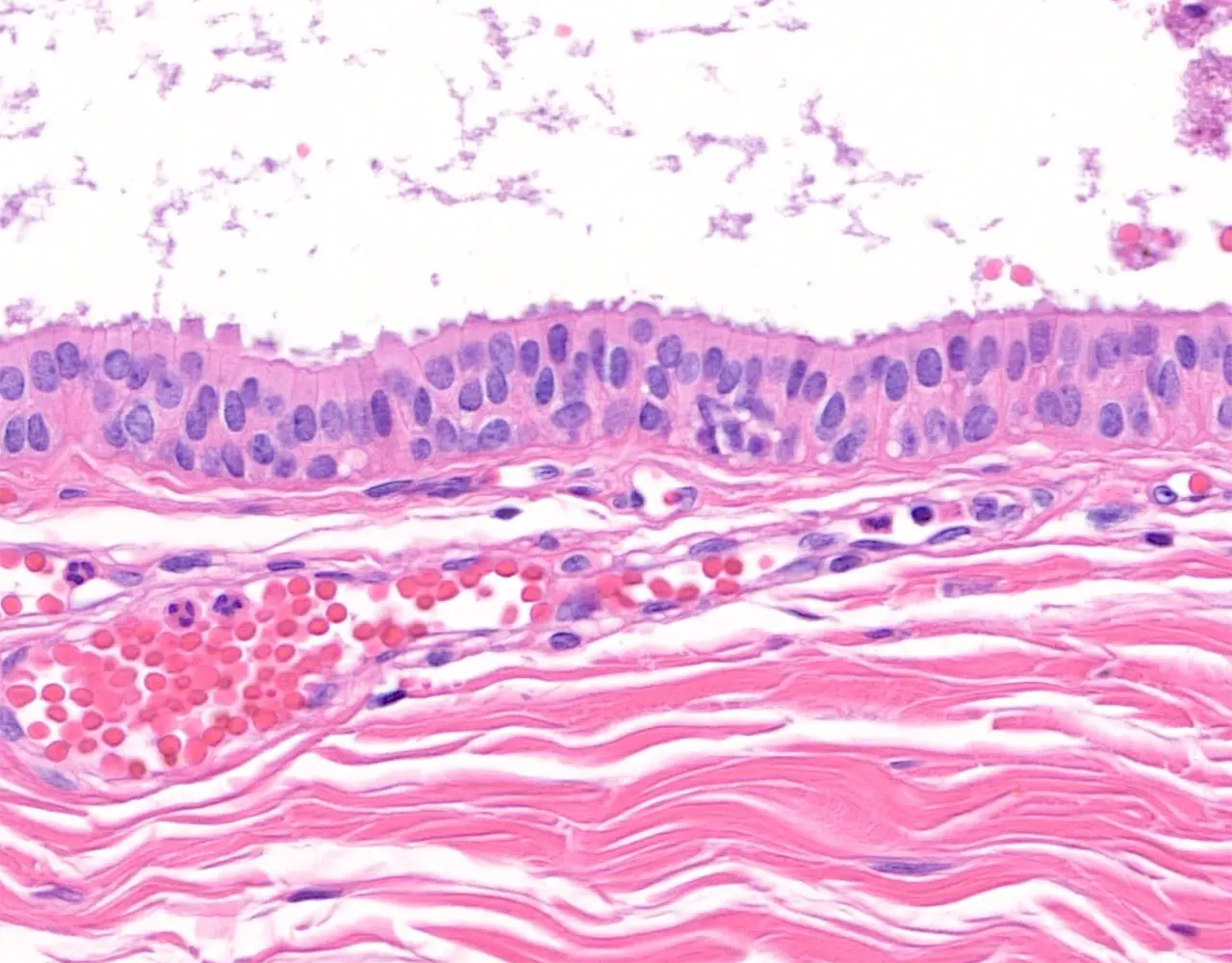
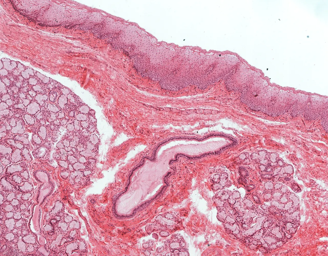
Interlobular and Main Excretory Ducts :
- They are larger than intralobular ducts and are located within the connective tissue septa.
- Their epithelium may be simple columnar, pseudostratified columnar, or stratified columnar.
- Near the oral cavity, the epithelium may become stratified squamous.
- Goblet cells may be present.
Distinctive Features
-
Parotid Gland :
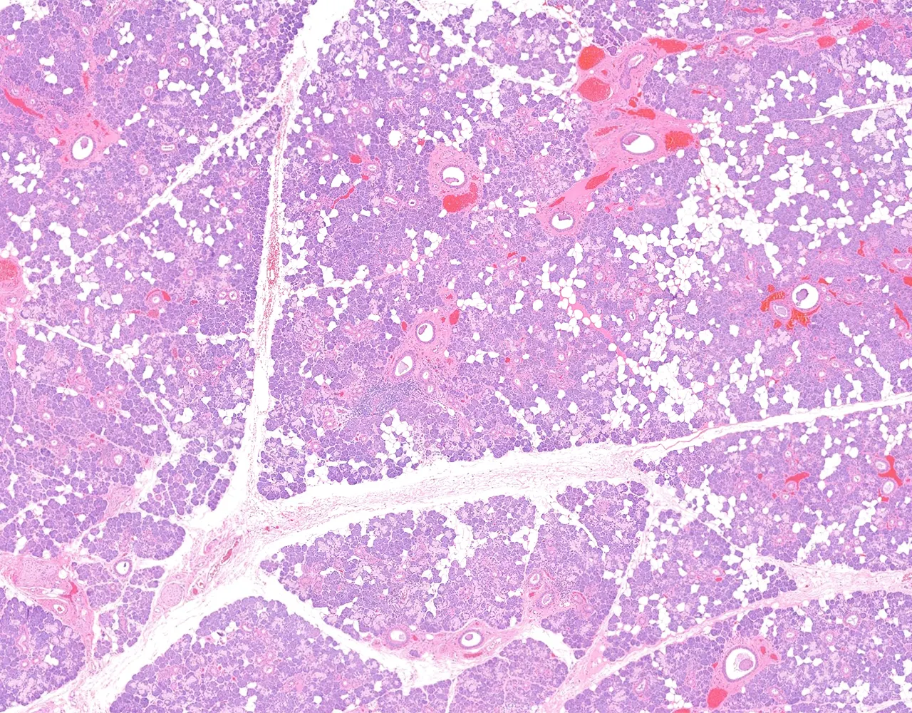
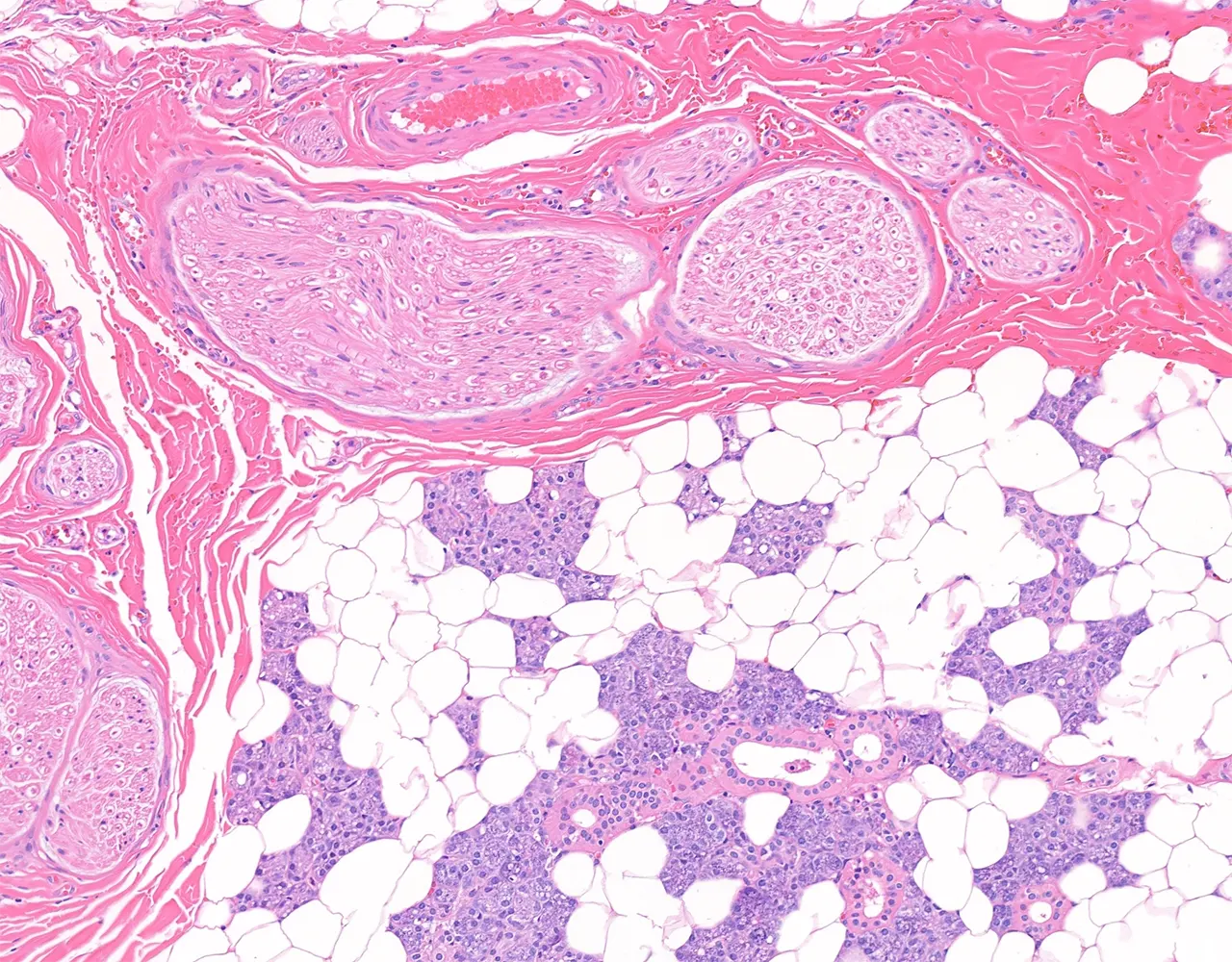
- The parotid gland is composed almost entirely of serous acini.
- It often contains abundant mature adipose tissue, especially in adults.
- Large nerve bundles, including branches of the facial nerve, may be seen within the gland.
- Intraparenchymal lymph nodes may be present and may contain benign salivary gland inclusions.
- Occasional sebaceous cells may be observed.
-
Submandibular Gland :
- The submandibular gland contains less adipose tissue than the parotid gland.
- It is a mixed seromucous gland composed mainly of serous acini, with fewer mucous acini.
- Striated ducts are usually prominent.
-
Sublingual Gland :
- The sublingual gland is predominantly mucous, and serous demilunes may be seen around mucous acini.
- Intercalated ducts and striated ducts are usually short and less conspicuous than in the parotid and submandibular glands.